When people think of an ICU, they picture around-the-clock critical care for individuals experiencing life-threatening scenarios. At Pet ER, we believe that pets deserve the same high level of critical care. That’s why Pet ER is proud to serve as a dedicated ICU to East Setauket pets and those in our neighbouring communities.
Whether your cat needs extensive oxygen therapy or your dog requires life-saving intervention, our team of seasoned and patient centered veterinary clinicians is here for them. Although this can be a stressful time for any pet owner, we hope you breathe a little easier knowing that your beloved companion is in good hands.
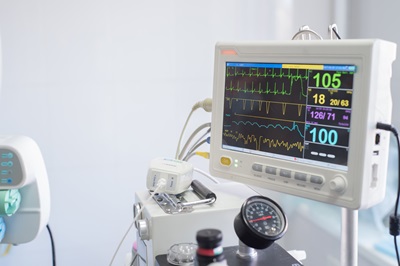
Yes. Our specialized ICU services include monitoring and caring for your pet overnight. Although you can’t be there for your pet, our team will work around the clock to keep them in stable condition.
Although there are various components that are similar between emergency care and what’s offered in an ICU, they are different. For example, you would rely on an ICU when your pet is experiencing a life-threatening emergency and when they’re stable and on the road to recovery but still require consistent monitoring.